Smart phone and gemology
Smartphone & Gemmology
by Gagan Choudhary
We, as gemmologists or buyers often feel the need of portable equipment which can assist us in identifying a gem especially during field trips – at mines, in gem markets, trade shows, etc. Although there have been portable kits containing folding polariscope, dichroscope, etc available in the market, a simple and innovative approach could be the use of our mobile phones in collecting the information provided by a polariscope or a dichroscope. The introduction of smartphones has enabled us to carry out various tasks with a single gadget – from making simple phone calls to messaging to exploring the internet to photography and much more – these have become an integral part of our lives. These smartphones can also put to use in gemmological applications!
We are basically looking at two parts of a smartphone – the digital camera and the display screen. Few top model phones have such good quality cameras that they compete some of the point-and-shoot digital cameras. Even mid to low end smartphones can take some usable images if taken with patience and experience, under proper lighting conditions. Today, the display screen used in a smartphone has a wide range with different technologies, some of them are – TFT LCD, IPS-LCD, Resistive Touchscreen LCD, Capacitive Touchscreen LCD, OLED, AMOLED, Super AMOLED, Retina Display, Haptic / Tactile touchscreen and Gorilla Glass, details of which can be viewed here. Most LCD (liquid crystal display) screens are sources of plane-polarised light; therefore, the screen can be used to check the optic character as well as the pleochrosim of a gem. However, it is important to first check the polarisation of the screen using a polarising filter or even polarising sunglasses. Such hand-held polarising filer or polarising sunglasses are used to make the smartphone screen, a polariscope. In the author’s experience, the earlier generation LCD screens provide the best results but something like Capacitive Touchscreen LCD also gives extremely satisfying and conclusive results. This is to be kept in mind that the screen colour should be white for gemmological applications.
This article outlines the various uses of a smartphone in gemmological applications and how a smartphone with some basic accessories can assist in identifying a gem or taking good-quality photographs and photomicrographs.
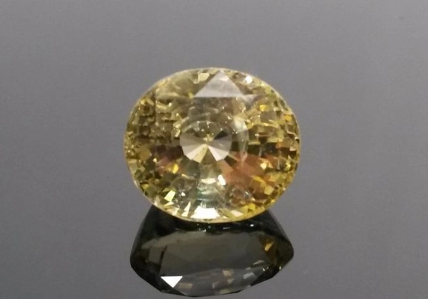
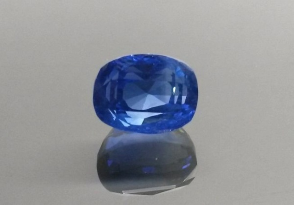
Here are few examples of images taken from the author’s smartphone camera.

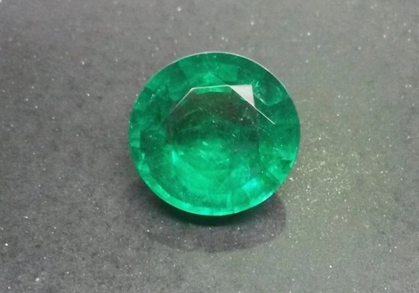
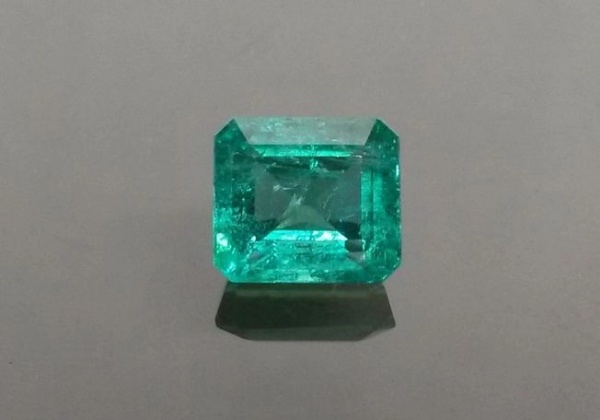
Judging optic character
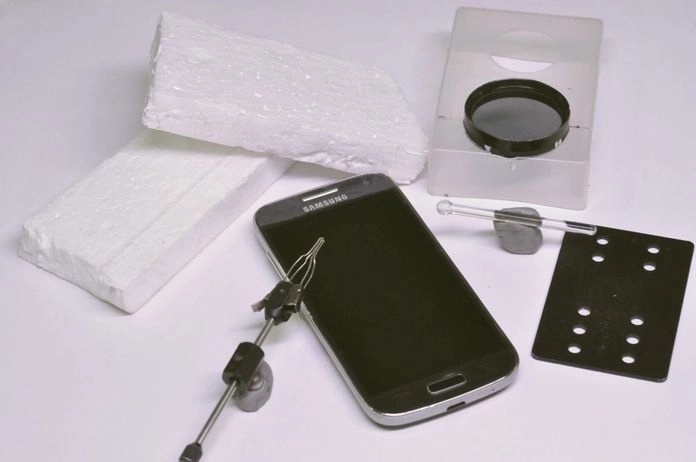
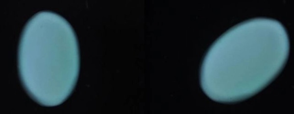
Optic character of a gem or mineral can be judged with the use of an additional polarising filter positioned perpendicular to the plane of polarisation of the light emitted by the screen. This can be done be viewing and rotating the polarising filter against the screen, and fixing the polariser at the position where the screen appears darkest – this position is the cross-polarised position of a polariscope. The author fixed the polariser on a plastic box as illustrated in figure 1 to keep the hands free. This set up now can be used as a polariscope – place the stone on the phone screen table down and rotate as done in a polariscope and note the reaction. Results obtained are illustrated in figure 3.

Resolving optic figures
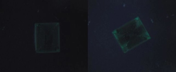
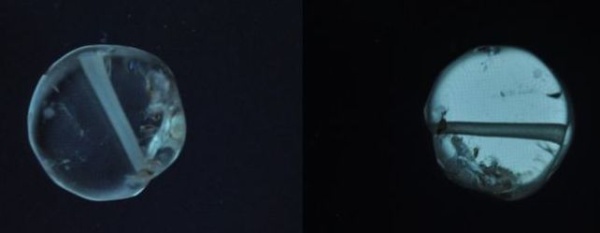
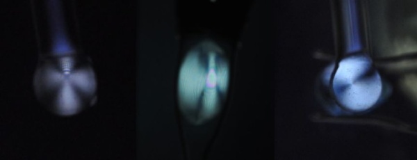
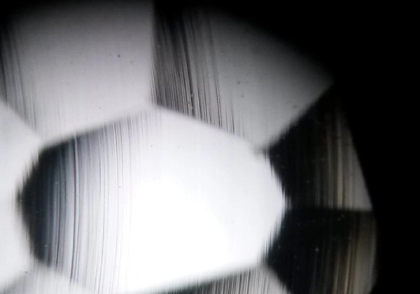
The same set-up can also be used to resolve optic figures using a conoscope once anisotropic nature is established. Following are few examples of optic figures resolved in smartphone-polariscope.
Judging pleochroism
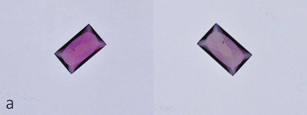
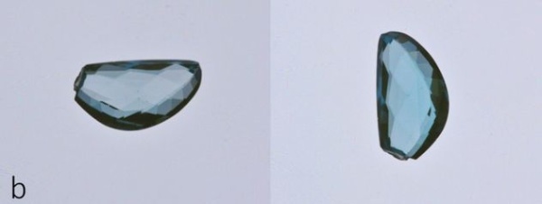
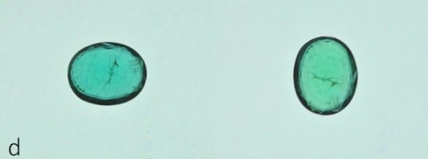
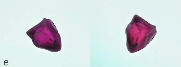
While judging pleochroism, no set up is required other than the white screen. Simply place the stone table down on the screen and observe the shift in colour while rotating it; the colour shift will be typically seen when rotated 90 degrees as illustrated in figure 5.

Taking photomicrographs
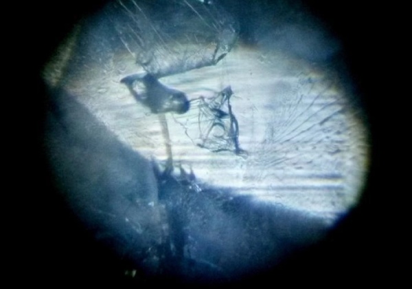
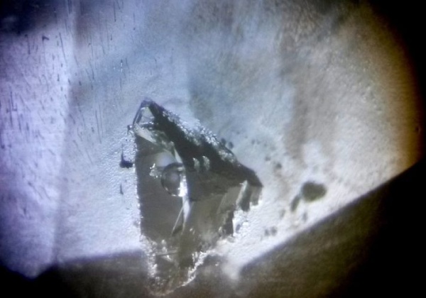
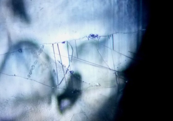
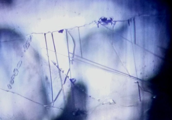
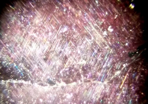
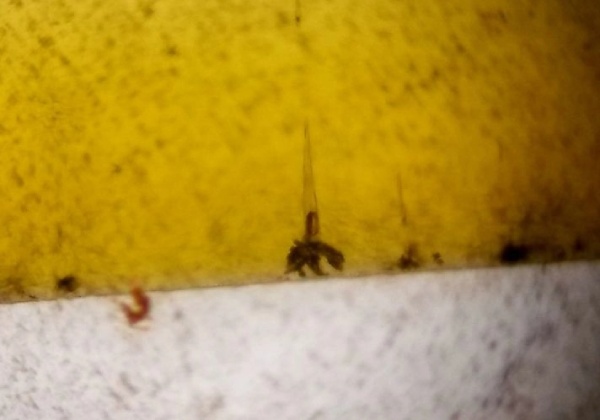
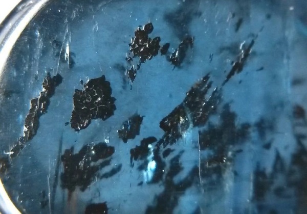
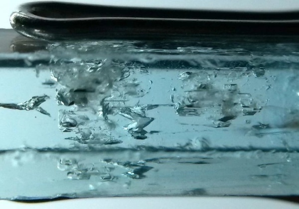
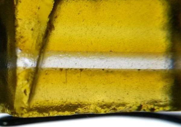
The author managed to take photomicrographs through a microscope as well as a 10x loupe. It is a basic criteria for any microscopy technique to properly orient and focus the inclusion, starting with the lowest magnification, and then increasing the magnification as per the requirement. The smartphone camera is positioned at the centre of one of the oculars at a distance (~ 1 cm) where the image is visible on the screen. Keep in mind to hold the camera as steady as possible. Also, when magnification is changed, one need to readjust the focus, therefore it is important to first practice holding the phone with two hands and then only one, where the other hand can be used to focus the object. Once the image is visible on the screen, zoom option of the phone can also be tried out to get a smaller field of view. The author managed to get some good quality photomicrographs even at high magnifications. Here are few examples:

Taking photomicrographs using a 10x loupe
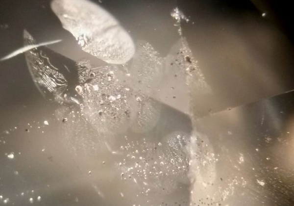
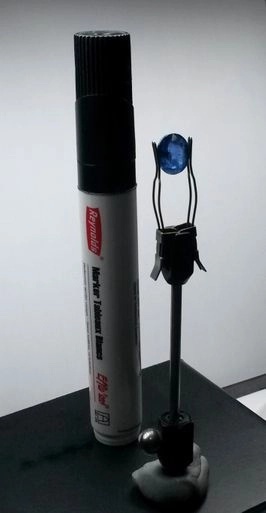
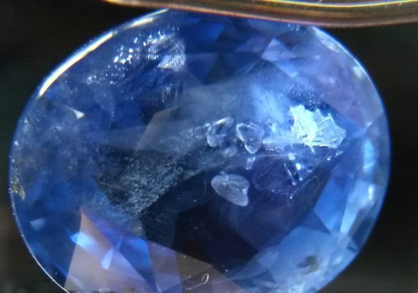
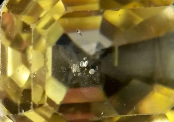
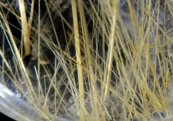
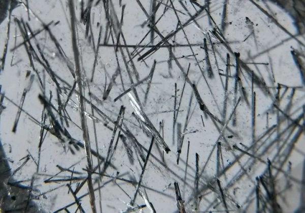
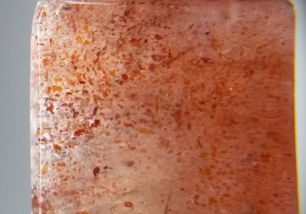
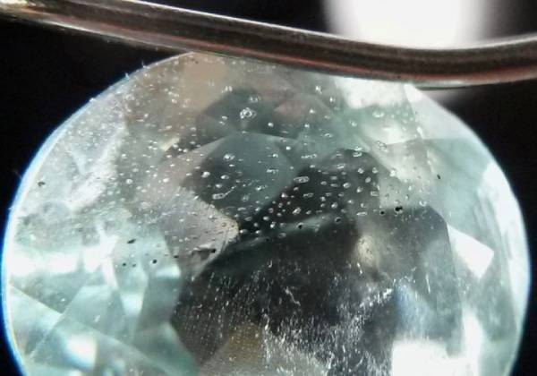
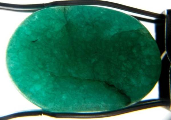
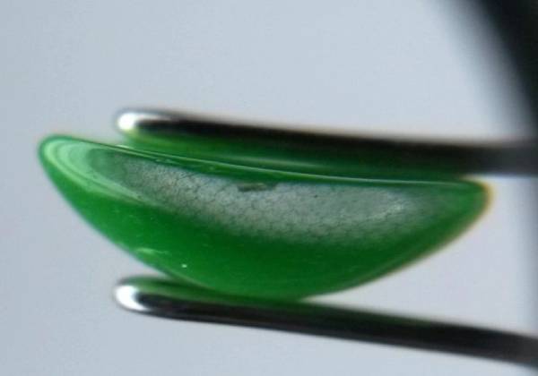
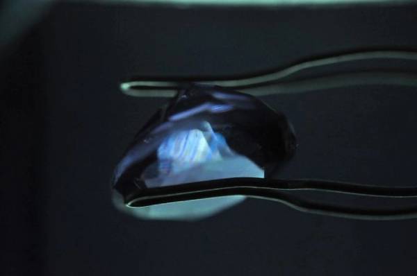
Although having the drawback of limited magnification, some good quality photomicrographs can still be captured using a simple 10x loupe, and can prove to be very useful arsenal for those who want to keep a check on the stones given for approvals. The microscope provides the luxury of using various types of illuminations, which cannot be replicated exactly during observations with a loupe. However, dark-field loupes are available in the market, but the author used the standard loupe and placed a black object behind the stone, to get the dark-field illumination (figure 14). Such set up provided reasonably good quality images, but again important factor is the correct distance between the object and the loupe to get the accurate focal point; this can also be done by playing around with the camera zoom. The best images were produced when the loupe was held closest to the camera lens.
Following are few examples of images captured through a 10x loupe and a smartphone camera….
Conclusions
Macro-photography attachments are however available in the market for some models of phone cameras, but good-quality images can be captured without these also; one has to keep in mind the stability of the camera, proper focussing and illumination. Although, smartphone cameras cannot replace the SLR cameras in terms of professional quality images, but they definitely have the ability to capture workable images. In addition, the phones’ display screen has an added advantage of turning it into a makeshift polariscope-cum-dichroscope, with great portability.
References
Boehm E. (2014) Photomicrography using a smartphone camera. The Journal of Gemmology, Vol. 34, No.1, pp 6-7
Devouard B., Bornet R., Notari .F, Rondeau B., and Fritsch E. (2010) Use your LCD screen as a gemological tool. Gems & Gemology, Vol. 46, No. 4, pp 325-326
Overton T.W. (2010) Smartphone photomicrography. Gems & Gemology, Vol. 46, No. 4, pp 325-326
http://www.quickonlinetips.com/archives/2011/02/types-of-displays-touchscreens-in-smartphones/
All photographs and photomicrographs by Gagan Choudhary
Visit us at Online Courses to know more.
Visit us at Online Courses to know more.
Visit us at Online Admissions Open
Call Us On +91 8879026633 / 7400497744 / 022-42906666 Courses to know more.
trending post


